Leg Bones Diagram / Skeletal System Diagrams - The bones of the leg are the femur, tibia, fibula and patella.. The foot bones shown in this diagram are the talus, navicular, cuneiform, cuboid, metatarsals. Includes leg (femur, tibia, patella, and fibula) and foot (tarsals and digits) bones. Click now to learn more about the bones, muscles, and soft tissues tibia: Your leg bones are the longest and strongest bones in your body. These simple labelled diagrams of the bones of the lower legs and feet and the bones of the arms and hands this diagram shows the skeletal structure of the leg (anterior view) and foot (dorsal view).
Human anatomy diagrams show internal. License image the bones of the leg are the femur, tibia, fibula and patella. Its lower end helps create the knee joint. The bones of the leg are the femur, tibia, fibula and patella. These simple labelled diagrams of the bones of the lower legs and feet and the bones of the arms and hands this diagram shows the skeletal structure of the leg (anterior view) and foot (dorsal view).
Forever Horses: Anatomy of the Equine Hindleg from 2.bp.blogspot.com Cheek bone (zygoma) upper jaw (maxilla). Upper leg bones diagram using this technique dr brown measured the length of each giraffe s neck upper and lower leg radius and metacarpal bones and ankle phalanx bone figure 1 f i g u r e 1. These simple labelled diagrams of the bones of the lower legs and feet and the bones of the arms and hands this diagram shows the skeletal structure of the leg (anterior view) and foot (dorsal view). High quality realistic skeleton legs. Lower jaw (mandible) collar bone. Learn vocabulary, terms and more with flashcards, games and other study tools. You'll learn about the muscles, bones, and other structures of each area of the leg. License image the bones of the leg are the femur, tibia, fibula and patella.
Each leg is made up of four bones.
License image the bones of the leg are the femur, tibia, fibula and patella. Includes leg (femur, tibia, patella, and fibula) and foot (tarsals and digits) bones. Start learning with free skeleton diagrams, bone labeling exercises and skeletal system quizzes. The bones of the leg are the femur, tibia, fibula and patella. These simple labelled diagrams of the bones of the lower legs and feet and the bones of the arms and hands this diagram shows the skeletal structure of the leg (anterior view) and foot (dorsal view). Your leg bones are the longest and strongest bones in your body. 8 bones (femur, patella, tibia, and fibula). Download 2,401 bones diagram stock illustrations, vectors & clipart for free or amazingly low rates! At the same time, the bones and joints of the leg and foot must be strong enough to support the body's weight while remaining flexible enough for movement and balance. Most bones (particularly the long bones of the arms and legs — which make up the appendicular skeleton) have a hard outer shell known as cortical bone. Bones in the human bodies and names. The foot bones shown in this diagram are the talus, navicular, cuneiform, cuboid, metatarsals. They allow you to move and provide support for your upper body.
These simple labelled diagrams of the bones of the lower legs and feet and the bones of the arms and hands this diagram shows the skeletal structure of the leg (anterior view) and foot (dorsal view). Health diagram bone skeleton leg knee science anchor chart human human body. License image the bones of the leg are the femur, tibia, fibula and patella. Start learning with free skeleton diagrams, bone labeling exercises and skeletal system quizzes. Click now to learn more about the bones, muscles, and soft tissues tibia:
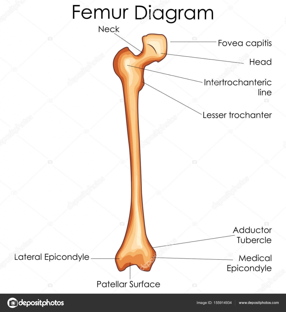
Femur bone diagram | Medical Education Chart of Biology ... from st3.depositphotos.com Your leg bones are the longest and strongest bones in your body. Most bones (particularly the long bones of the arms and legs — which make up the appendicular skeleton) have a hard outer shell known as cortical bone. The bones involved in it, however, are only the femur and the tibia, although the smaller bone of the leg, the fibula, is carried along in the movements of flexion, extension, and slight rotation that this joint. The bones of the leg are the femur, tibia, fibula and patella. 3d general characteristics model 3d human leg bone: Start learning with free skeleton diagrams, bone labeling exercises and skeletal system quizzes. The foot bones shown in this diagram are the talus, navicular, cuneiform, cuboid, metatarsals and calcaneus. Download 2,401 bones diagram stock illustrations, vectors & clipart for free or amazingly low rates!
The foot bones shown in this diagram are the talus, navicular, cuneiform, cuboid, metatarsals and calcaneus.
At the same time, the bones and joints of the leg and foot must be strong enough to support the body's weight while remaining flexible enough for movement and balance. When you stand or walk, all the weight of your upper body rests on them. You'll learn about the muscles, bones, and other structures of each area of the leg. They allow you to move and provide support for your upper body. The bones of the leg are the femur, tibia, fibula and patella. At the microscopic level, this hard outer. Bones of the leg and foot, lower leg bone anatomy, leg bones anatomy, leg muscles, leg bones diagram, leg bone structure, leg anatomy muscles, parts of the lower leg. The largest and most medial leg bone, forming both the knee and ankle joints. Upper leg bones diagram using this technique dr brown measured the length of each giraffe s neck upper and lower leg radius and metacarpal bones and ankle phalanx bone figure 1 f i g u r e 1. The foot bones shown in this diagram are the talus, navicular, cuneiform, cuboid, metatarsals and calcaneus. Start learning with free skeleton diagrams, bone labeling exercises and skeletal system quizzes. The tibia is the main bone of the leg, forming what is more commonly known as the shin. The bones involved in it, however, are only the femur and the tibia, although the smaller bone of the leg, the fibula, is carried along in the movements of flexion, extension, and slight rotation that this joint.
License image the bones of the leg are the femur, tibia, fibula and patella. You'll learn about the muscles, bones, and other structures of each area of the leg. The bones involved in it, however, are only the femur and the tibia, although the smaller bone of the leg, the fibula, is carried along in the movements of flexion, extension, and slight rotation that this joint. They allow you to move and provide support for your upper body. Lower jaw (mandible) collar bone.
The Leg Skeleton | Anatomy bones, Human body anatomy, Leg ... from i.pinimg.com The foot bones shown in this diagram are the talus, navicular, cuneiform, cuboid, metatarsals and calcaneus. New users enjoy 60% off. Includes leg (femur, tibia, patella, and fibula) and foot (tarsals and digits) bones. Your legs are two of your most important body parts. The foot bones shown in this diagram are the talus, navicular, cuneiform, cuboid, metatarsals and calcaneus. The foot bones shown in this diagram are the talus, navicular, cuneiform, cuboid, metatarsals. Cheek bone (zygoma) upper jaw (maxilla). Learn how to draw the femur, patella, tibia, and fibula in this lesson!
Health diagram bone skeleton leg knee science anchor chart human human body.
The human leg, in the general word sense, is the entire lower limb of the human body, including the foot, thigh and even the hip or gluteal region. At the microscopic level, this hard outer. Bones in the human bodies and names. High quality realistic skeleton legs. Most bones (particularly the long bones of the arms and legs — which make up the appendicular skeleton) have a hard outer shell known as cortical bone. Human anatomy diagrams show internal. Start learning with free skeleton diagrams, bone labeling exercises and skeletal system quizzes. They allow you to move and provide support for your upper body. The tibia is the main bone of the leg, forming what is more commonly known as the shin. The foot bones shown in this diagram are the talus, navicular, cuneiform, cuboid, metatarsals. Health diagram bone skeleton leg knee science anchor chart human human body. The foot bones shown in this diagram are the talus, navicular, cuneiform, cuboid, metatarsals and calcaneus. The bones of the leg are the femur, tibia, fibula and patella.